Cruciate Rupture
It is one of 2 ligaments that cross inside the stifle (veterinary term for canine knee) joint. These ligaments prevent the femur and tibia from sliding back and forth on each other.
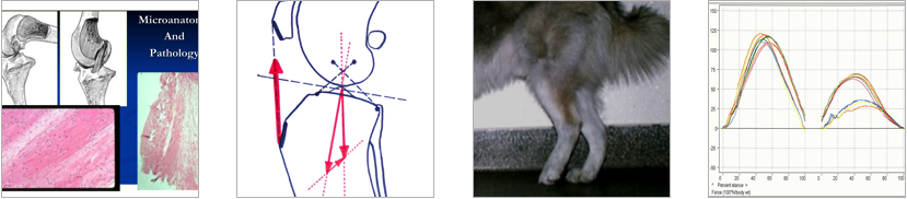
These ligaments are especially important for our canine and feline companions because their hind limbs are flexed (bent) most of the time. When they put weight on their hind legs, the tendency is for the femur to roll or slide back and down the top of the tibia. The CrCL stops this downward slide, and, because their legs are usually bent, these ligaments are under constant stress and strain.
The biomechanical stress strains the ligament which leads to fraying, partial and complete tear. There are other factors that can cause or exacerbate this issue, like obesity, poor diet, joint inflammation, limb deformities, poor or extreme exercise, and a number of other genetic factors. The combination of factors that cause Cruciate Rupture is part of why this is often referred to as Canine Cranial Cruciate Disease.
Due to the frequency of the injury and need for a solution, the veterinary profession has focused tremendous energy on this problem.
If you suspect that your companion has suffered a CrCL injury, familiarize yourself with the common signs and make an appointment with your veterinarian for a proper examination.
Signs
- – A history of mild lameness in the same limb that seems to come and go before this sudden worsening of symptoms.
- – Sudden lameness on one rear limb to a degree that weight can not be borne.
Lameness includes:
-
- Pain
-
- Decreased range of motion
-
- Loss of muscle mass (muscle atrophy)
-
- Abnormal posture when standing, getting up, lying down, or sitting
-
- Abnormal gait when walking, trotting, climbing stairs, or turning
-
- Nervous system signs — confusion, trembling, etc.
-
- Swelling and inflammation
- Grating or grinding on joint movement
Cruciate rupture can manifest as an acute/sudden lameness or a gradual deterioration, so if you suspect that your companion is experiencing a form of hind limb lameness, contact your veterinarian and schedule a physical exam.
Also, be aware that, while CrCL rupture is the most common culprit, sometimes the primary cause of lameness is somewhere else… hips, ankle, neurological, etc.
Consequences
In addition to pain, here are some other consequences of CrCL rupture:
-
- – Ligament deterioration releases a combination of inflammatory factors from the ligament.
– Increasing instability of the joint from the weakened ligament causes arthritis to develop quickly within the joint.
– Every time the pet bears weight on the affected leg, the femur slides down the tibial plateau with nothing to halt its movement. This sliding action damages a cartilage cushion in the joint called the meniscus. Once the meniscus is torn arthritic change accelerates and perceived pain worsens.
– Weight bearing studies have shown that dogs with a damaged CrCL bear only about 20-30% of the normal amount of weight on the affected leg. As a result, the weight is shifted to the other rear leg placing even more stress on that limb’s CrCL.
Treatments
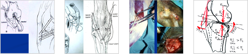
Numerous procedures have been used to stabilize the “drawer sign” in the joint. These procedures include, but are not limited to: lateral suture, lateral fabellar suture, over-the-top, Tibial Plateau Leveling Osteotomy (TPLO), Triple Tibial Osteotomy (TTO), and Tibial Tuberosity Advancement (TTA).
Once CrCL is confirmed, your veterinarian will discuss the different options they offer and provide you with referral information so that you can consult a Specialist Surgeon.
Innovations
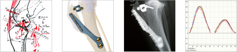
Slobodan Tepic, Dr. Sci., Dipl. Ing., Founder & CEO of KYON, and Prof. Pierre M. Montavon, Head of Small Animal Surgery at the School of Veterinary Medicine, University of Zurich, invented and developed the Tibial Tuberosity Advancement (TTA) procedure to restore joint stability in cranial cruciate deficient canines. Their research showed that if the angle between the patellar tendon and the tibial plateau could be reduced to a perpendicular (90 degree) angle, then the joint force would be shifted so that the joint is stable even without the CrCL.
The TTA involves an osteotomy of the tibia, just behind the tibial tuberosity, hence the name. The tibial tuberosity is advanced to align the patellar tendon to 90 degrees to the tibial plateau. This new alignment eliminates the need for a CrCL and results in a stable joint.
The advanced tibial tuberosity is secured using titanium implants specifically designed and manufactured by KYON to meet the unique demands of the procedure, providing a sound biological fixation*. A bone graft is often packed in the open area of the osteotomy to speed up healing, which takes approximately 8 weeks.
KYON launched TTA for cranial cruciate deficiency in dogs in early 2004, following three years of clinical testing and implant development/refinement. While positive publications on the technique and short-term clinical outcomes are encouraging, recent in vitro studies on the restoration of normal contact mechanics and stifle kinematics show that TTA may mitigate the progression of osteoarthritis. Our belief is that the most important benefits and advantages of TTA are expected in long-term, trouble-free performance. read publications on Tibial Tuberosity Advancement>>
KYON’s TTA procedure has been used in over 100,000 cases by more than 950 surgeons and has become an important addition to the canine cranial cruciate repair armamentarium.
*The rapid acceptance of TTA has motivated several other companies to offer similar implants. KYON has neither collaborated with nor endorsed any of the competitors in the manufacture, promotion or sale of their TTA systems. KYON Veterinary Surgical Products is the only authorized distributor of KYON products in the United States. Search our website Surgeon Database to find a surgeon performing KYON procedures with KYON implants.Your companion deserves the best.
Testimonials

Talu after TTA site has healed
Breed: American Pitbull Terrier
Date of Surgery: 11-06-2012
Procedure: Right TTA, MPL repair and arthroscopic meniscal evaluation
Surgeon: Dr. Geoffrey Hutchinson, DVM, MS, DACVS
Clinic: Boundary Bay Veterinary Specialty Hospital (BBVSH)
Referring Veterinarian: Dr. Lomond, Paws and Claws Animal Hospital
Comments from owner:
‘We are SO pleased with Talu’s recovery from his combined TTA and patella luxation repair! Dr. Hutchinson did a fantastic job and we would highly recommend BBVSH to all pet lovers! We adopted Talu, a 3 yr old Pitbull from LAPS, knowing he had issues with his right hind leg. After having him home for only 4 months, he became non weight bearing on that leg. After x-rays and consultations we decided to go ahead with surgery to give him his mobility back. We were really impressed by how much time Dr. Hutchinson took to explain the procedures to us. The follow up visits post surgery continued to show us how professional and helpful all the staff is at BBVSH. Talu still limped from time to time, but improved with every month post-op. After 6 months he was medication free and bounding around with wild abandon in true Pitbull style! In November 2013, one full year after surgery, Talu is happy, healthy and has no limitations to his exercise. He can enjoy life to the fullest, romping with other dogs and hiking with us, on a regular basis. We can’t thank Dr. Hutchinson enough for helping Talu! – Elaine and Greg
More photos of Talu:

Talu after TTA site has healed

Talu after TTA site has healed

Frankie and friends
Breed: American Bulldog Cross (female)
Date of Surgery #1: 05/07/2013
Procedure: left TTA
Date of Surgery #2: 05/22/2013
Procedure: right TTA
Surgeon: Dr. Geoffrey Hutchinson, DVM, MS, DACVS
Clinic: Boundary Bay Veterinary Specialty Hospital (BBVSH)
Referring Veterinarian: Dr. Ashley Danyluk, Shaughnessy Veterinary Hospital
More about Frankie
This is ‘Frankie’, a beautiful American Bulldog cross who came to BBVSH one year ago due to lameness in both hind legs. She was diagnosed with a left cranial cruciate tear AND a right partial cranial cruciate ligament tear by Dr. Geoff Hutchinson. Frankie’s owner Sharon opted to proceed with the Kyon Tibial Tuberosity Advancement (TTA) procedures as recommended by Dr. Hutchinson. The left limb was showing greater lameness so it was the left hind limb that was treated first. At the 2 week recheck Frankie was doing so well that the right limb was also treated with the Kyon TTA procedure. Sharon followed the strict confinement instructions given by our surgery department and is now enjoying life to the fullest!
Comments from owner:
“I just wanted to say it’s been a year since Frankie had her surgeries on her knees and all I ever wanted was to see her enjoy life again. Thanks to your skill and care, she is back to being her old self or perhaps a bit bionic!!! Here is a picture of her taken today enjoying a romp in the woods.
Frankie and I both say thank you” – Sharon H.
More photos of Frankie:

Frankie in the creek

Frankie 1 year post-op